
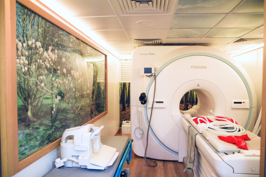
Magnetic Resonance Imaging
With Magnetic Resonance Imaging (MRI), a number of body parts can be examined, including the brain, spine, eye sockets, joints, and soft tissues. People frequently undergo MRI scans, which are also performed frequently at Movement Referrals. In contrast to X-rays, MRI makes use of magnetic fields instead of radiation.
An MRI scan involves obtaining many images of the patient as slices or cross-sections. In comparison to conventional X-rays, these slices allow individual structures to be viewed directly without being obstructed by other structures. In order to get the best information about any particular structure, the slice direction can be changed.
At Movement Referrals, we have an MRI scanner with a strong magnetic field (1.5 tesla, so-called a ‘high field’ scanner), allowing us to take good quality images, assisting our Specialists in diagnosing the patient, and enabling the scans to be taken relatively quickly.
Despite the fact that high field scanners can perform scans more quickly than low field scanners, each of the multiple sequences (components of the scan) takes approximately five minutes to complete, and the patient must remain perfectly still throughout all of them. This results in MRI scans being performed under general anaesthesia (unlike in humans). It is necessary to use MRI-compatible non-magnetic anaesthesia monitoring equipment (which shows the patient’s blood pressure, breathing and blood oxygen levels, etc.) as MRI scanners have large magnets as their main component.
Diagnostic imaging with MRI provides excellent detail and contrast of soft tissues (in contrast to radiography). Due to the lack of interference from surrounding air or bones, magnetic imaging can reveal exquisite detail in areas that are otherwise difficult to image such as the brain and spinal cord (the nerves that run from the brain to the spine).

CT-Scan
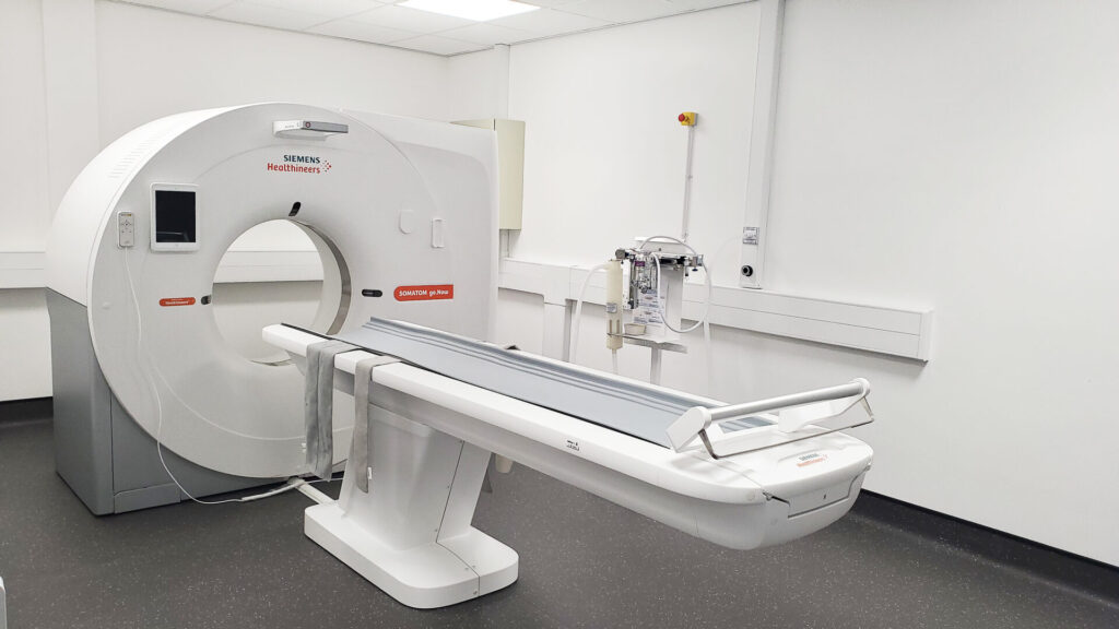
The term CT refers to computed tomography. The technique uses X-rays to reconstruct cross-sectional images of an animal in a scanner using a computer. A CT scan gives more details about bones (skull, spine, joints) compared to an MRI scan, but less detail about soft tissues like brain and spinal cord. Therefore, this technique is often used to image the vertebral column and can, on occasion, be used in conjunction with MRI to provide us with more information about your pet’s condition.
A myelogram is the procedure of injecting a dye into the fluid surrounding the spinal cord in order to take an X-ray of the spinal cord. The dye allows X-rays to clearly show the shape of the spinal cord. Slipped discs can change the shape of the spinal cord and are frequently diagnosed using myelography. An MRI scan allows your veterinarian to visualise spinal cord tissue itself in addition to providing accurate information on the spinal cord’s shape (and whether pressure is present). A CT-myelogram, however, combines injecting dye with a CT to get cross-sectional images that are more detailed.