
Background

The menisci (plural of meniscus) are important structures in the knee joint of dogs (and most mammals), playing a pivotal role in distributing forces across the bone surfaces, and contributing to joint stability. Their position is illustrated here on the right. They are comprised of fibrous cartilage, with the main collagen fibres arranged from front to back (illustrated with grey lines in the picture).
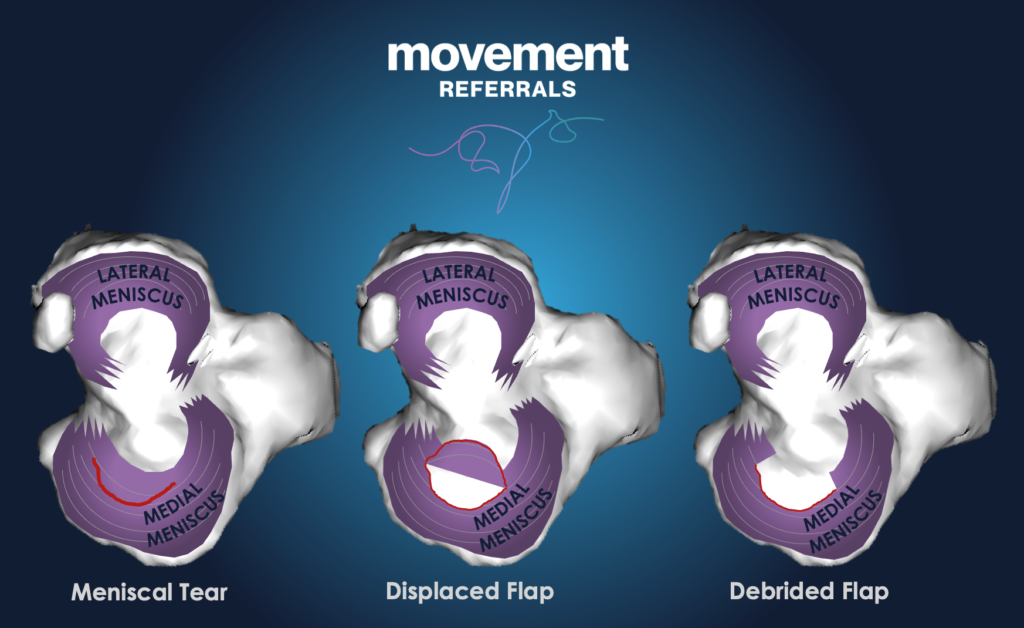
Meniscal injuries on their own are uncommon in dogs, but the medial meniscus is very prone to tearing when the cranial cruciate ligament is ruptured. The most common type of tear is the “bucket handle”: the medial meniscus is torn along the line of its main fibres, creating a “flap” with attachments at the front and the back as shown in the picture on the left.
Clinical Signs
When meniscal injuries have occurred before CCL failure is diagnosed, we say they are “concomitant”. Dogs with CCL failure and concomitant meniscal injury might be more lame than those without. Sometimes, meniscal injuries occur at some point after CCL failure has been surgically treated: we call these “late” meniscal injuries. In these cases, the dog might have been recovering well, and then become more lame again.
Some dogs with meniscal injury will have an audible “click” when they walk, believed to be caused by an unstable flap flicking on its remaining attachments.
Diagnosis
Certain findings on clinical examination can raise suspicion for meniscal injury, including a click or a clunk on manipulation, pain on the medial aspect of the affected knee, and pain on flexion of the knee.
An important part of diagnosing meniscal injuries in post-surgical patients is to rule out other causes of lameness and knee pain, such as infection or implant failure. Radiographs and the collection and analysis of joint fluid are helpful in this regard, but meniscal injuries cannot be confirmed with these methods.
Although meniscal injuries can be identified using MRI or CT scans, this is very rarely done in dogs as both methods require sedation or general anaesthetic, and can use up financial resources. Therefore, most cases of suspected meniscal injury are confirmed surgically, which means they can be treated at the same time. Arthroscopy offers a minimally-invasive surgical option for inspecting the menisci <Video 1>.
Treatment
If a meniscal injury is causing enough pain and lameness that it is noticeable to the owner, recovery and final outcome can usually be improved with surgical debridement. For “bucket handle” tears, the flap’s attachments are cut to release the unstable portion and allow removal.
Expected Outcome
Most dogs respond well to debridement of meniscal injuries, and lameness generally improves within a few days to weeks. In the longer term, dogs with meniscal injuries might develop more osteoarthritis in the joint. This is the result of instability, and greater pressure in some of areas
Publications from Publications from Movement Vets surgeons on meniscal problems
- Wustefeld-Janssens BG, Pettitt RA, Cowderoy EC, Walton, MB, Comerford EJ, Maddox TW, Innes JF Peak Vertical Force and Vertical Impulse in Dogs With Cranial Cruciate Ligament Rupture and Meniscal Injury. Veterinary Surgery, 45(1), 60-65. https://doi.org/10.1111/vsu.12419