

The shoulder joint in dogs is composed of the joint between the scapula (shoulder blade) and humerus. Dogs do not have a clavicle (collar bone) and cats only have a small, vestigial collar bone.
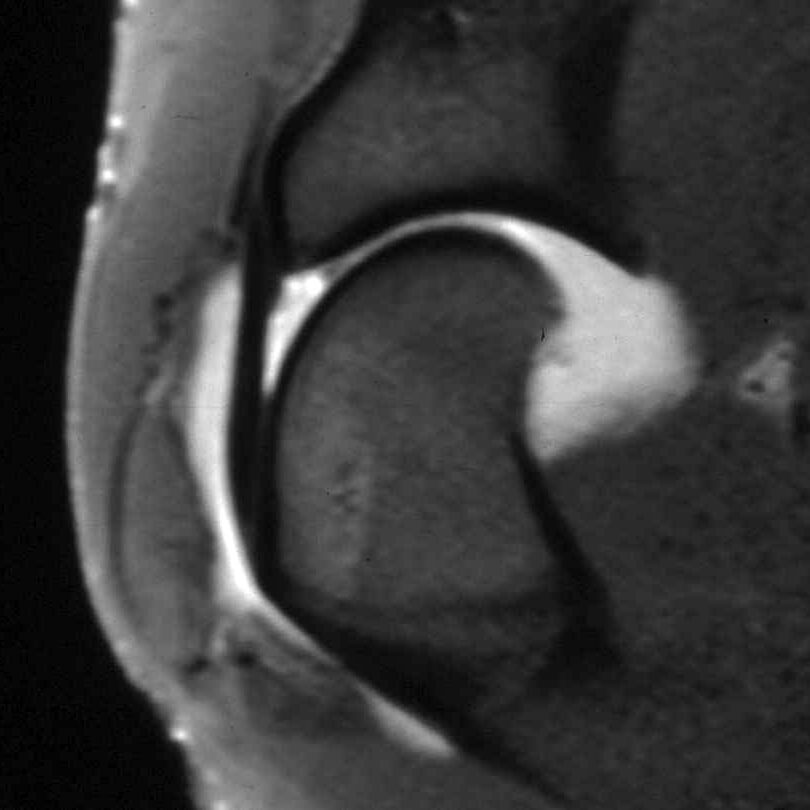
Because the shoulder joint is surrounded by soft tissues, plain X-rays of the shoulder can be rather unrewarding, particularly in adult dogs with shoulder lameness. In these instances, we might use advanced imaging techniques such as MRI or arthroscopy.
The shoulder joint can be affected by a variety of conditions and some of the more common ones are discussed here:
Osteochondritis dissecans (OCD)
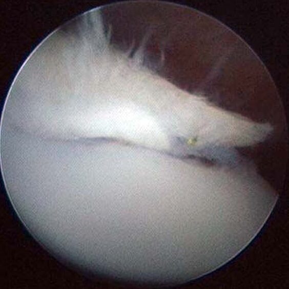
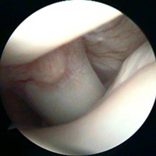
Osteochondritis dissecans is a developmental disorder of larger breed dogs that typically starts to cause signs at 5-7 months of age, but sometimes later. Breeds affected by OCD include giant breeds such as Great Danes and popular breeds such as Labrador and Golden Retrievers, as well as Border Collies. The cause is not clear but it is more common in male dogs. It represents a failure of the joint surface to develop correctly and the cartilage, which lines the joint surface, can become partially detached from the underlying bone; this causes pain and because the cartilage is typically partially detached, the underlying tissue cannot heal.
Recommended treatment for OCD that is causing persistent pain is surgical removal of the loose cartilage and, in our opinion, this is best performed with keyhole surgery (arthroscopy). Arthroscopy minimises trauma to the soft tissues and allows for a rapid recovery. In addition, arthroscopy provides the surgeon with magnification and access to all parts of the joint.
Soft tissue injuries to the shoulder joint
Working dogs and agility dogs are at increased risk of injuries to the tendons, ligaments and muscles around the shoulder joint. Whilst muscular injuries should heal with an appropriate period of rest, tendon and ligament injuries can be more problematic and persistent. Accurate diagnosis of these injuries can be challenging and most will not be apparent on plain X-rays. Careful clinical examination may provide useful information but advanced imaging techniques such as arthroscopy or MRI may be required to identify the exact cause. Treatment for these injuries can vary depending on the exact nature of the problem.
Osteoarthritis of the shoulder joint
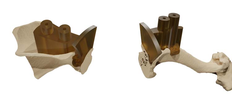
Osteoarthritis of the shoulder joint can be commonly noted on X-rays of older dogs. However, the majority of dogs seem to cope well with mild osteoarthritis. Occasionally shoulder osteoarthritis is associated with persistent pain and medication may be required. If this is not sufficient, fusion (arthrodesis) of the shoulder can be a helpful procedure. Arthrodesis of the shoulder needs careful planning and 3D modelling of the bones is very useful to allow for accurate cutting of bones and precise contouring of bone plates prior to surgery.
Functional outcomes after shoulder arthrodesis can be very good.

Publications from Movement Vets surgeons on shoulder problems
1. Pettitt RA, Innes JF: Arthroscopic management of a lateral glenohumeral ligament rupture in two dogs. Veterinary and Comparative Orthopaedics and Traumatology 21:302-306, 2008.
2. O’Neill T, Innes JF: Treatment of shoulder instability caused by medial glenohumeral ligament rupture with thermal capsulorrhaphy. Journal of Small Animal Practice 45:521-524, 2004.
3. Innes JF, Brown G: Rupture of the biceps brachii tendon sheath in two dogs. Journal of Small Animal Practice 45:25-28, 2004.
4. Mitchell RAS, Innes JF: Lateral glenohumeral ligament rupture in three dogs. Journal of Small Animal Practice 41:511-514, 2000.