
Basic facts regarding growth in puppies and kittens
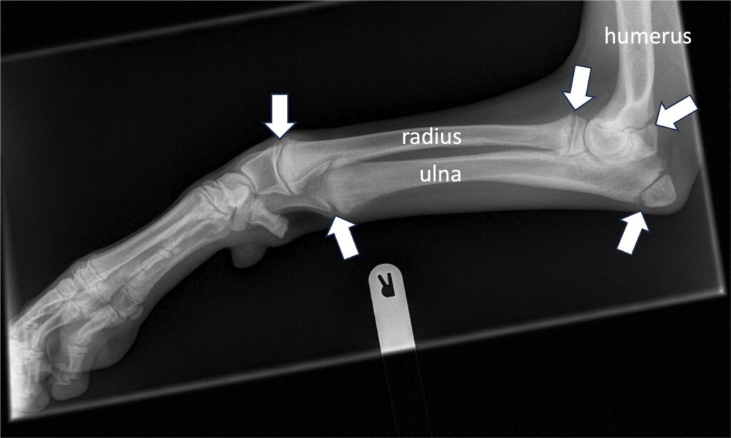
Dogs grow remarkably quickly. If one thinks that a large or giant breed dog is fully grown by 12-18 months of age, compared to a human being, that is a remarkable growth velocity. When vertebrate animals develop, their bones first form as cartilage models, which turn to bone through a process called ossification. Bones gain some of their length from growth at the very ends, but most of it develops at the “growth plate” (also called physes). Cartilage forms to widen the growth plate, and then turns to bone in a “wave” of ossification behind the growing part. Growth plates must grow at the same rate across the growth plate, otherwise the bone might start to deform. In addition, in the antebrachium (the forearm), there are two long bones, the radius and ulna, and so these two bones must grow at the same rate, otherwise there can be bowing of the antebrachium. Not surprisingly, the antebrachium is therefore the most common site of growth deformities in puppies.
Sometimes, there are disturbances in these processes that effect how the bone develops. These disturbances might be due to injury, metabolic abnormalities, infection, or something else.
Growth disturbance can result in various deformities, including:
- Limb length discrepancy, when one or more legs ends up being too short.
- Angular and torsional deformities, when one or more limbs end up being bent or twisted
- Joint incongruity, when one or more of the joints end up not fitting together well.
Some growth abnormalities are best left untreated, but many of them require surgical intervention to optimise the chance of having good function in adulthood. The most difficult aspect of managing growth abnormalities, in our opinion, is decision making about if, when, and how to intervene.
Keywords
- Growth-plate (also called a physis): The zone of cartilage near the ends of a growing bone from where most of the length is gained.
- Lameness: limping.
- Radius and Ulna: the two bones in the forelimb between the elbow and the carpus (wrist).
- Osteotomy: a surgical cut through a bone.
- Ostectomy: the surgical removal of a section of bone.
Causes
- Breed-related deformities. Some breeds of dog look the way they do because they have bent or twisted bones. These breeds are called “chondrodystrophic”, and examples include Shih-Tzu, Lhasa-Apso, Dachshunds, French Bull Dogs and Spaniels. Usually, the shape of their legs does not obviously affect them, and they function normally. Sometimes, though, the legs might grow to be “excessively” bent and/ or twisted, and this can cause lameness.
- Injury. Growth plates are particularly susceptible to compression injury. Sometimes, there will be pain, lameness or swelling that comes on suddenly, and growth plate injury is identified early on. Other times, there might be no, mild, or temporary signs at the time of the injury, and growth abnormalities become evident weeks or months later.
- Nutrition. Overfeeding or underfeeding or underfeeding growing animals can affect their skeletal development, and this is probably most important in giant breed dogs who grow very quickly.
- Environment and exercise. Environment has the biggest impact on skeletal development in the first few weeks of life.
- Other. Less common causes of growth disturbances include conditions that cause inflammation of the growth plates or bones (eg, bacterial physitis, metaphyseal osteopathy), or other problems with growth plate development (eg retained cartilaginous cores)
Clinical Signs
- Lameness/ limping. Sometimes, pain and/ or lameness is the only clinical sign, and the limb might look relatively normal. This can occur when there is joint incongruity because the bones have not developed to match each other. This usually occurs in the elbow joint when the radius has grown more than the ulna, or vice-versa.
- Stumbling/ tripping. If the shape of the legs is very abnormal, they cannot take the load normally when the animal is walking or trotting, and this can look like they are tripping over.
- Cosmetic. Legs might look obviously bent or twisted.
Investigation
- The most important part of the assessment is the clinical examination, as this helps to determine whether bone shape, joint incongruity, or laxity or tension in the soft tissues are the main consideration.
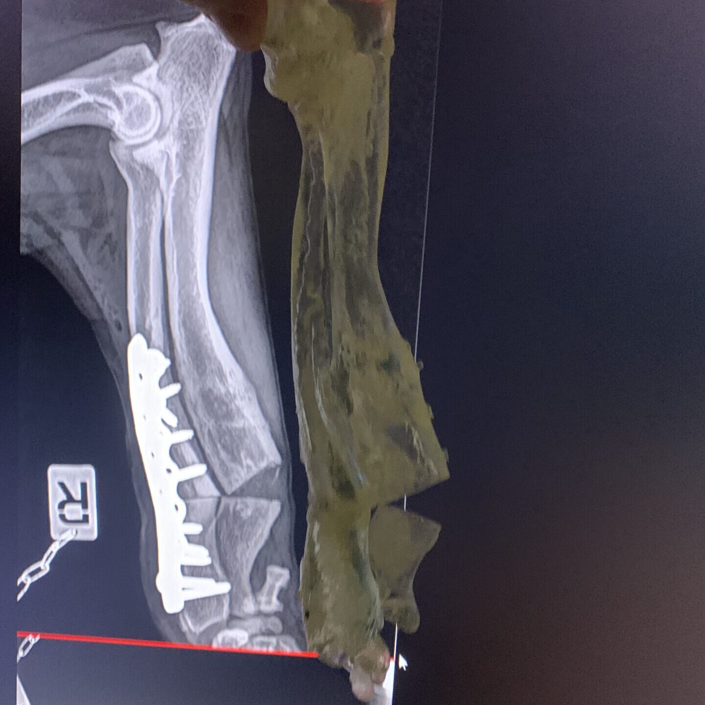
- Radiographs (X-rays) can be helpful to assess for angular deformities (bends) and limb-length discrepancies. They are less useful for quantifying torsional deformities (twists), and are less sensitive than CT for detecting joint incongruity. Basic surgical planning can be performed on radiographs.
- CT scanning can give high-resolution, three-dimensional images of the bones and joints. It is a powerful tool for detecting and measuring all types of bone deformity and joint incongruity, and for advanced surgical planning.
Treatment
- Non-surgical treatment. Some growth deformities should be left untreated, especially if they are not causing any lameness or other mobility impairment.
- “Dynamic” ulna osteotomies/ ostectomy. In some cases, a cut through one of the two of bones in the forelimb between the elbow and the wrist can be enough to improve the congruity in the elbow joint.
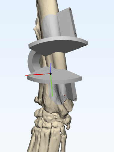
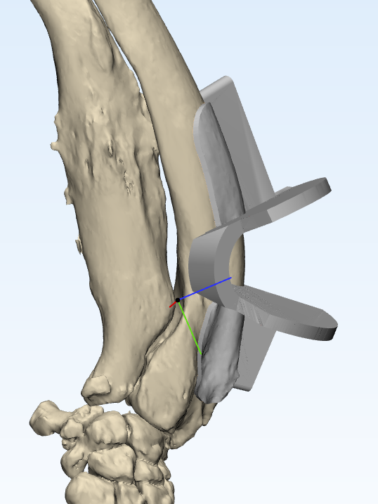
- Corrective osteotomies. It might be necessary to reduce the bends and/ or twists in the bone to improve limb function. This is generally done by cutting the effected bones, adjusting their shape, and fixing them in place while they heal. We might advise having patient-specific custom surgical guides made. We would usually aim to stabilise the bones with internal implants (e.g. bone plates) to make the post-operative care easier.
- Distraction osteogenesis. In rare cases, usually where there is still considerable growth potential in a bone, it might be advised to fit an adjustable external skeletal fixator. This is a frame around the leg that is attached to the bones with pins and wires, and can be adjusted in increments to gradually influence the shape and length of an affected bone.
Premature closure of antebrachial growth plates
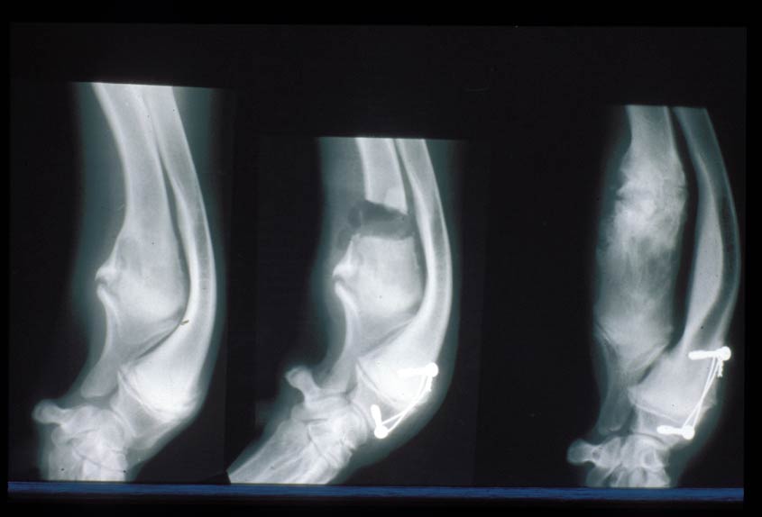
The most common site for growth deformities is the antebrachium. This is because the radius and ulna must grow at the same rate. 85% of the growth of the ulna comes from the distal growth plate and so it is not surprising that the most common growth problem is premature closure of the distal ulnar growth plate. This causes the ulna to act as a ‘bow string’ on the radius and the radius can start to bow (“radius curvus”). Treatment of this problem can take various forms often depending on the age of the puppy. Careful evaluation, planning and monitoring is necessary for best outcomes. If the problem is caught early, sometimes it is possible to influence growth to reduce the deformity. In other cases, it may be necessary to perform corrective surgery to acutely correct the deformity, and in other cases a gradual correction may be performed using external frames and pins.
Problems with the radial growth plates can also occur but are less common. The distal radial growth plate accounts for approximately 60% of the growth of the radius and the remaining 30% comes from the proximal growth plate. Loss of growth of the radius can cause ‘short radius syndrome’ which can lead to subluxation of the elbow joint. Asymmetrical disease of the distal radial growth plate can cause deformity of the limb (e.g. carpal varus).
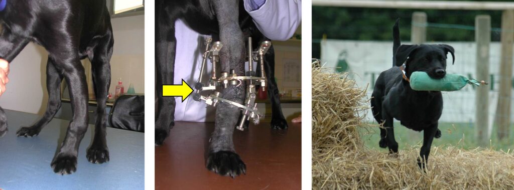
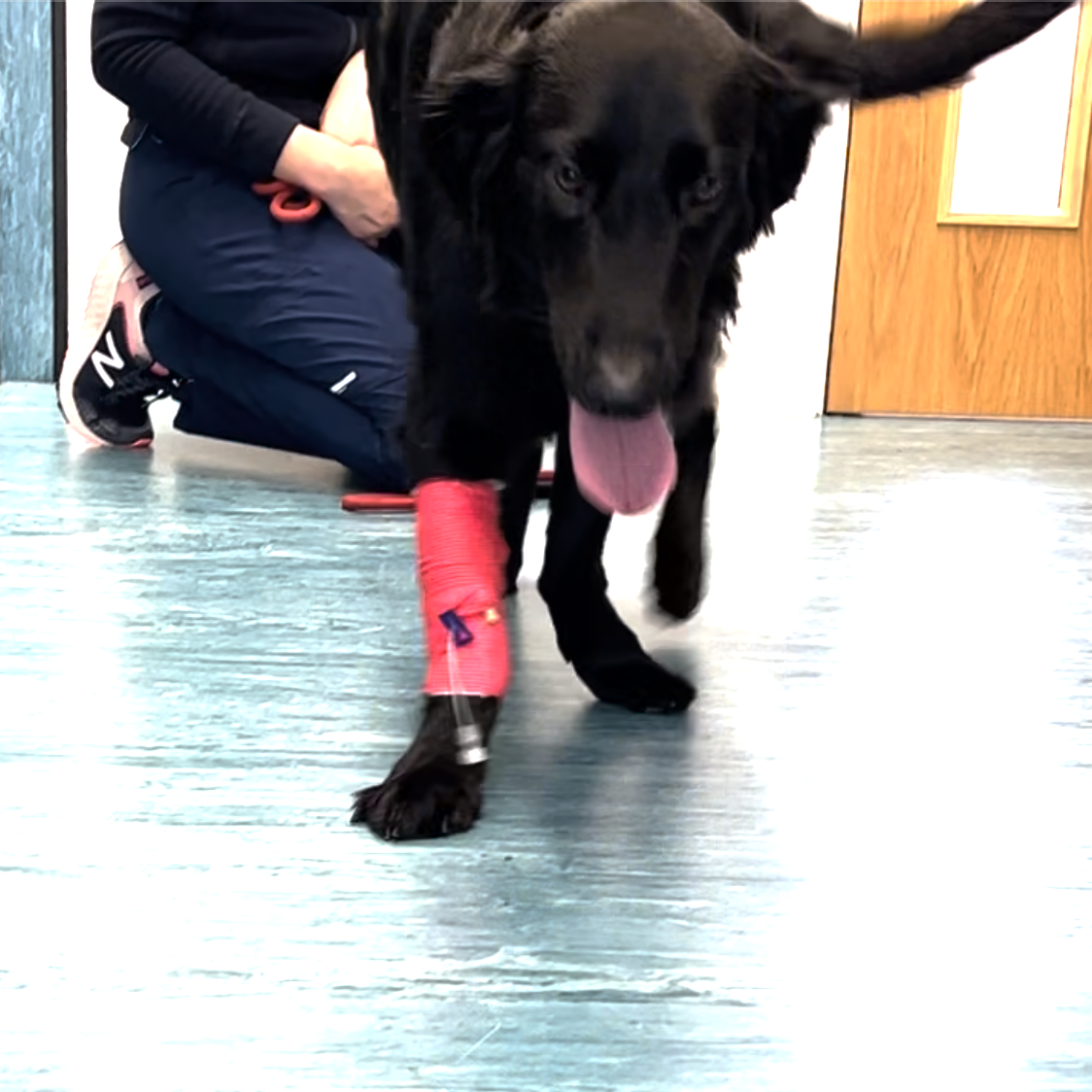
A Case of Growth Deformity Treated by Movement Referrals
The pictures here are of a Flat-Coat Retriever who had growth deformities in both legs.
Publications from Movement Vets surgeons
Innes, J. F., W. M. McKee, R. A. S. Mitchell, B. D. X. Lascelles, and K. A. Johnson. 2001. ‘Surgical reconstruction of ectrodactyly deformity in four dogs’, Veterinary and Comparative Orthopaedics and Traumatology, 14: 201-09.