
The elbow joint
The elbow joint is a complex joint made up of the humerus which articulates with both the radius and the ulna. As well as acting as a hinge, there is considerable rotationary movement within the elbow joint to allow for pronation and supination.
In dogs, the elbow joint can be a common seat of lameness problems, as well as fractures. Lameness issues are most commonly caused by coronoid disease (also called ‘elbow dysplasia’) although developmental conditions such as OCD and ununited anconeal process occur from time to time. The common fractures of the elbow joint are thought to be stress fractures and particular breeds are predisposed.
Coronoid disease (‘elbow dysplasia’)
Disease of the medial coronoid process is a poorly understood condition which most commonly presents in young (6-12 months) dogs of medium-large breed dogs (e.g. Labrador Retrievers, Bernese Mountain Dogs). However, it can also appear in adult dogs and in some smaller breeds. Necrosis and fragmentation of the medial coronoid process can occur, and this is associated with the initiation of osteoarthritis. Osteoarthritis may be mild or severe and can be a cause of pain in itself, above and beyond the coronoid disease.
Diagnosis of coronoid disease can be challenging because plain X-rays usually do not show the condition, although they may show some osteoarthritic changes. Imaging with a CT scan or viewing the interior of the joint with arthroscopy are much more sensitive methods to detect and assess the problem.
Because of the complexity of coronoid disease and elbow osteoarthritis, careful assessment of each individual case, against a backdrop of specialist, evidence-based knowledge, is preferred.

Osteochondritis dissecans (OCD)
Osteochondritis dissecans is a less common developmental disorder of larger breed dogs that typically starts to cause signs at 5-9 months of age, but sometimes later. Approximately 1% of young dogs with painful elbows have OCD. It represents a failure of the joint surface to develop correctly and the cartilage on the medial aspect of the distal humerus (the medial condyle), which lines the joint surface, can become partially detached from the underlying bone; this causes pain and because the cartilage is typically partially detached, the underlying tissue cannot heal.
Recommended treatment for OCD that is causing persistent pain is surgical removal of the loose cartilage and, in our opinion, this is best performed with keyhole surgery (arthroscopy). Arthroscopy minimises trauma to the soft tissues and allows for a rapid recovery. In addition, arthroscopy provides the surgeon with magnification and access to all parts of the joint.
Ununited anconeal process
In growing dogs, the aonconeal process of the ulna should fuse to the remainder of the ulna by 22 weeks of age. In some dogs, this fusion fails and the anconeal process detaches and becomes loose in the joint. This can cause pain, inflammation and secondary osteoarthritis. The condition can occur sporadically in many larger breed dogs, but the most common breed to be affected is the German Shepherd Dog. If diagnosed early, surgical treatment can allow for the anconeus to fuse.
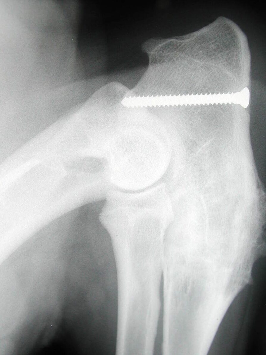
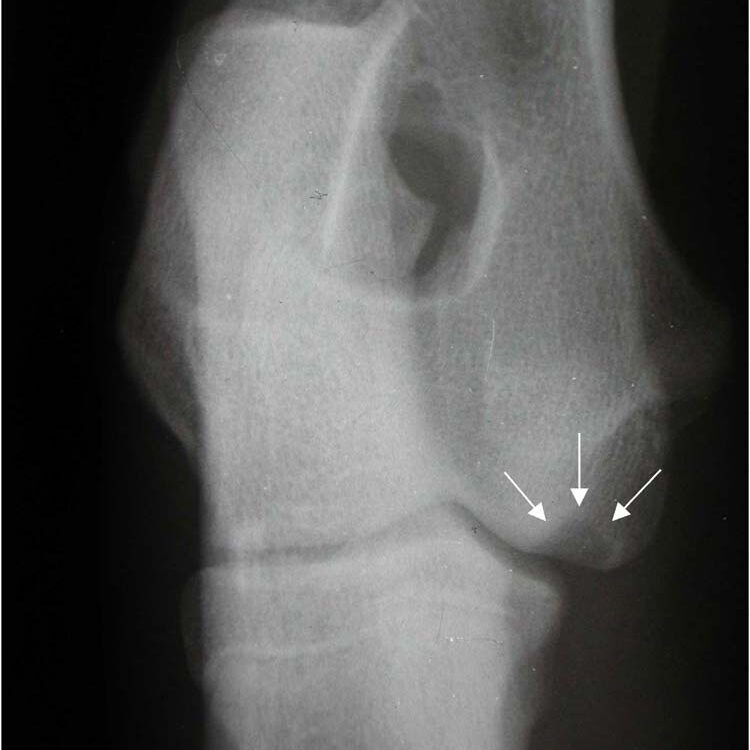
Humeral intracondylar fissure (HIF) (aka ‘incomplete ossification of the humeral condyle [IOHC])
Humeral intracondylar fissure is believed to be a stress fracture of the humeral condyle and is particularly prevalent in the English Springer Spaniel (ESS). Up to 14% of ESSs in UK have an abnormality within the humeral condyle which may indicate an increased risk for HIF or condylar fractures. HIF can cause chronic lameness and elbow pain and it can affect both elbows. Diagnosis is best made with CT scanning and, in most cases, surgical treatment is recommended.
Humeral intracondylar fissure is a particular interest of surgeons at Movement Vets. We have been at the forefront of the diagnosis and treatment of this condition for over 20 years. John Innes was co-author of the first publication to identify the problem in the UK, and Ben Walton has been working on methods to treat the condition for over a decade. Our recent publications have demonstrated improved success rates and reduced complication rates with the treatment methods we have developed. You can read more about Humeral intracondylar fissure here.
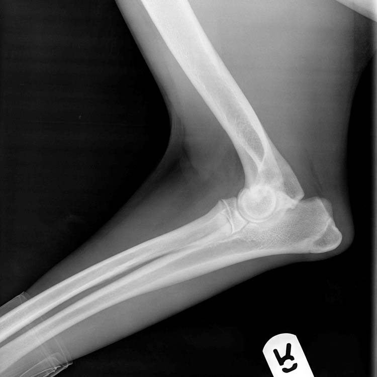
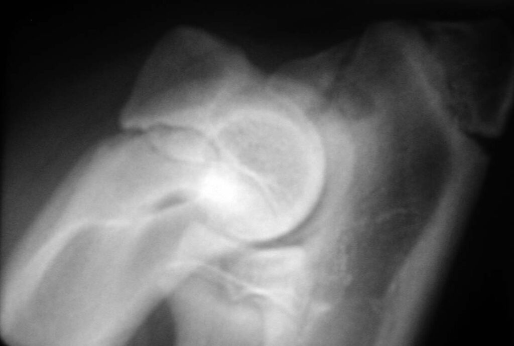
Fractures of the humeral condyle
Fractures of the humeral condyle are the most common fractures to occur in dogs. Because HIF can cause a weakness in the humeral condyle, these fractures can occur during normal activity. Because of the prevalence of HIF in the breed, English Springer Spaniels are prone to condylar fractures but other breeds such as French Bulldogs, Yorkshire Terriers and Labrador Rertrievers are also at increased risk
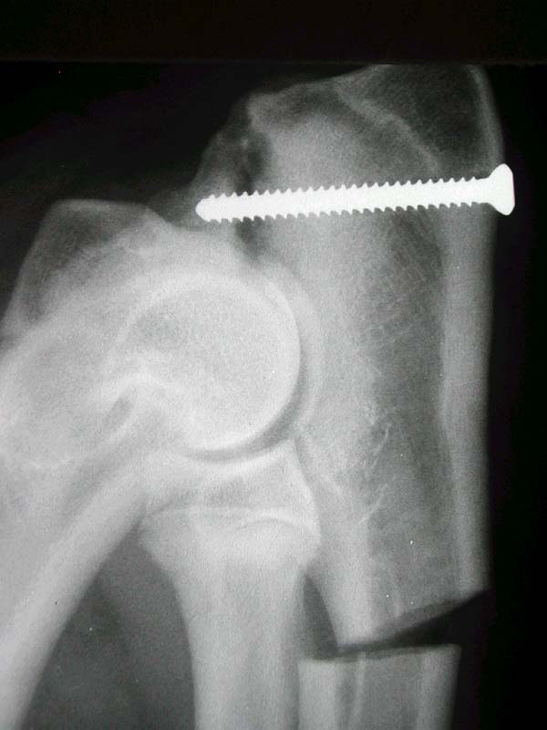
We have a particular interest in condylar fractures at Movement Vets. Ben Walton has worked with Fusion Implants to develop ‘anatomical plates’ which are specifically designed to treat these fractures in Springer Spaniels and French Bulldogs. These plates help the surgeon to accurately reduce the fractures and ensure that screws are in the optimal location. You can read more about our work on condylar fractures here.
Publications from Movement Vets surgeons on elbow problems
Butterworth, S. J. and J. F. Innes (2001). “Incomplete humeral condylar fractures in the dog.” Journal of Small Animal Practice 42(8): 394-398.
Lorenz, N. D., S. Channon, R. Pettitt, P. Smirthwaite and J. F. Innes (2015). “Ex vivo kinematic studies of a canine unlinked semi-constrained hybrid total elbow arthroplasty system.” Veterinary and Comparative Orthopaedics and Traumatology 28(1): 39-47.
McCarthy, J., E. J. Comerford, J. F. Innes and R. A. Pettitt (2020). “Elbow Arthrodesis Using a Medially Positioned Plate in 6 Dogs.” Veterinary and Comparative Orthopaedics and Traumatology 33(1): 51-58.
Pettitt, R. A., J. Tattersall, T. Gemmill, S. J. Butterworth, T. J. O’Neill, S. J. Langley-Hobbs, E. J. Comerford and J. F. Innes (2009). “Effect of surgical technique on radiographic fusion of the anconeus in the treatment of ununited anconeal process.” Journal of Small Animal Practice 50(10): 545-548.
Turner, B. M., R. H. Abercromby, J. Innes, W. M. McKee and M. G. Ness (1998). “Dynamic proximal ulnar osteotomy for the treatment of ununited anconeal process in 17 dogs.” Veterinary and Comparative Orthopaedics and Traumatology 11(2): 76-79.
Walton, M. B., E. Crystal, S. Morrison, J. Onyett, J. McClement, R. Allan, M. Straw and J. F. Innes (2020). “A humeral intracondylar repair system for the management of humeral intracondylar fissure and humeral condylar fracture.” Journal of Small Animal Practice 61(12): 757-765.